Medical imaging can look like a machine-centered field from the outside.
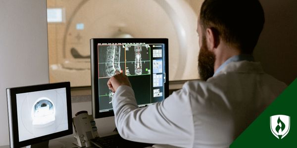
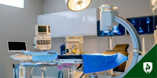
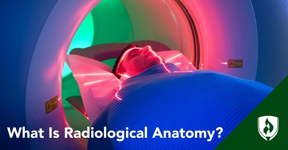
You've got magnetic resonance imaging (MRI) machines, X-ray machines, computed tomography, mammogram machines, mobile imaging units, digital images, monitors and specialized equipment that help care teams see what is happening inside the body.
But radiologic technology also depends on a very human skill: understanding the body well enough to capture images that are clear, useful and safe.
Radiologic technologists operate imaging equipment and produce diagnostic images, while radiologists are physicians who interpret those images and make diagnoses.1
That means a radiologic technologist’s work is all about getting the best images possible. And that involves knowing what anatomy needs to be shown, how the patient should be positioned and whether the image is clear enough to support the next step in care.
Radiographic anatomy is the discipline of studying human anatomy using radiographic films (X-ray images)—getting used to how the body looks through the lens of an X-ray machine. But you have to have a solid foundation of human anatomy first.
That is where radiological anatomy comes in. Dawn McNeil, program director and instructor in Rasmussen University’s School of Health Sciences Radiologic Technology program, explains that students first need a foundation in anatomy and physiology before learning how those same body structures appear on radiographic images.
In other words, radiological anatomy helps connect textbook anatomy to real diagnostic imaging. It gives radiologic technologists the knowledge they need to understand what they are looking at, how to image it properly and how to support quality patient care.
If you are considering a career in radiologic technology, you'll need a thorough understanding of the human body—in both traditional and radiological anatomy.
What does radiological anatomy include?
Radiological anatomy is the study of how the body’s structures appear through medical imaging.
Instead of looking at anatomy only through textbook diagrams or models, radiologic technologists need to understand how bones, organs, air spaces and other structures appear on images created through X-rays, fluoroscopy, CT and other imaging tools.
“The student needs to know the full anatomy of body parts from a typical A&P (anatomy and physiology) course first, and then we teach them how those body parts appear on a radiographic image,” McNeil says.
That shift matters because anatomy does not always look the same on a medical image as it does in a textbook.
McNeil uses the chest as an example.
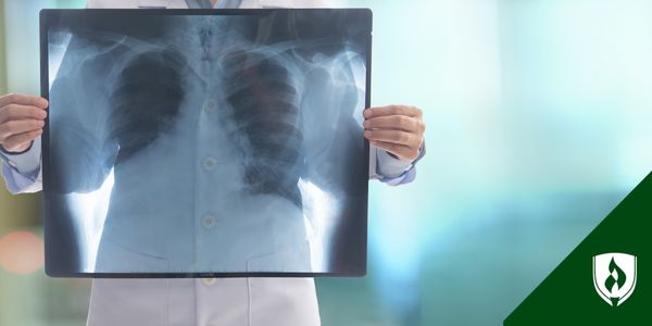
Students may first learn about the ribs, lungs, pleura, heart and major vessels in an anatomy and physiology course. Then, after a chest X-ray, they learn how those structures appear on a radiographic image: ribs appear brighter because they are made of bone, lungs appear darker because they are mostly air and the heart creates a recognizable shadow between the lungs.
Radiological anatomy also connects closely to radiographic positioning. Technologists need to understand what anatomy should be visible, how surrounding structures may overlap and whether the image was taken in the correct position. They may also need to consider whether the exposure factors were appropriate for producing a useful image.
That is why radiological anatomy is often taught alongside positioning, projections and image analysis. Rasmussen University’s Radiologic Technology program, for example, includes coursework in Radiologic Positioning and Anatomy covering areas such as the chest, bony thorax, upper extremities, abdomen, standard projections, positioning terminology and image analysis.2
Why radiological anatomy matters for radiologic technologists
Radiological anatomy is not just background science. It is part of the everyday work that helps radiologic technologists position patients, capture useful images, adapt exams and support patient safety.
It helps rad techs position patients accurately
Patient positioning is one of the clearest ways anatomy knowledge becomes practical. A technologist needs to understand not only which body part is being imaged, but also how position, angle and projection can change what appears on the final image.
For example, students may learn positioning terms such as AP, PA and lateral views. These terms describe how the body is positioned in relation to the X-ray beam and image receptor. The same body part can look different depending on where the X-ray enters, where it exits and what part of the body is closest to the image receptor.
And getting a clear, useful image is difficult. Imaging technique changes for every part of the body, every machine and if contrast agents like iodine, barium or gadolinium might be in use. Radiologic technologists can even specialize in certain machines, imaging modalities or fields, such as:
- Nuclear medicine, using position emission tomography (PET, scans), common in cancer treatment
- Mammography (breast cancer screenings and mammogram machines)
- Magnetic Resonance Imaging (MRI)
- Computed tomography scanners (CT/CAT scans)
- Note: Ultrasound/sonography is a separate branch of study, different from radiologic technology.
McNeil says students often need to adjust from colorful anatomy diagrams to radiographic images that appear in shades of gray. Working with a skeleton model can help them see how bones shift as body parts move into different positions, then compare those changes to the resulting image.
“The aha moment comes from using the skeleton model and then comparing it to the image,” McNeil says.
It supports diagnostic image quality
Radiologic technologists do not diagnose patients, but they are responsible for producing images that are useful for diagnosis. That means the image needs to show the correct anatomy clearly enough for the radiologist or provider to interpret.
Images that rad techs capture are a huge part of how physicians diagnose, how they plan surgeries and assess if treatment is working.
This is another reason radiological anatomy matters. Technologists need to recognize whether the correct body part is shown, whether the image was taken in the right position and whether the exposure factors were appropriate. Without that understanding, it would be harder to know whether an image is useful or whether adjustments are needed.
Rasmussen University’s Radiologic Technology program includes coursework in radiographic evaluation, image criteria, healthy and diseased images, equipment quality control and making appropriate adjustments as needed.3 These skills connect anatomy knowledge to the practical work of producing quality diagnostic images.
“Part of quality patient care in radiology is getting the image right the first time,” McNeil says.
It helps technologists adapt to real patients
Patients do not always fit the ideal setup shown in a textbook or lab demonstration. A patient may be injured, anxious, elderly, pediatric, in pain, unable to stand or unable to move into a standard position.
Rad techs might need to use their left hand at times, even if they're right-handed, or vis versa.
Radiological anatomy helps technologists adjust the exam while still capturing the needed anatomy. Students learn patient care and positioning skills, including how to assess patient sensitivity and condition to modify procedures while keeping patients informed.4
“For example, if a patient is holding their arm against their body and doesn’t want to move it for a standard forearm X-ray," McNeil says, "the technologist can rotate the X-ray tube to adjust the position, allowing the patient to remain comfortable.”
She gives another example of a standing abdominal image used to assess fluid levels. If the patient cannot stand, the technologist may use a different position, such as a decubitus position, to capture the needed image.
These adjustments require more than technical equipment skills. They require understanding what anatomy needs to appear in the image and how to capture it while accounting for the patient’s condition.
It contributes to patient safety and radiation protection
Radiological anatomy also supports radiation safety. A technologist who understands the area of interest can better position the patient, capture the correct view and reduce the need for repeat exposures.
Every X-ray exposure matters, McNeil emphasizes. Even when exposure is limited, repeating images adds radiation, which is why students practice positioning in the lab before working with patients in clinical settings.
“We can’t take an exposure for trial and error; we must know how to do it correctly,” McNeil says.
That is part of why anatomy, positioning, image quality and patient care are so closely connected in radiologic technology. The goal is not only to take an image, but to take the right image as safely and clearly as possible.
What parts of anatomy do radiologic technologists study?
Radiologic technologists study many parts of the body, but the goal is not to memorize anatomy in isolation.
They need to understand anatomy in a way that helps them position patients, capture the correct images and recognize whether the needed structures are visible.
Skeletal anatomy
Bones and joints are a major part of radiologic technology, and they are often what people think of first when they picture an X-ray. Radiologic technologists need to understand the spine, ribs, skull, pelvis, upper extremities, lower extremities and joints so they can capture images that show alignment, structure and the area of concern clearly.
Skeletal anatomy also connects closely to positioning. A small change in how a shoulder, wrist, knee or ankle is positioned can change what appears in the image. That is why students learn not only the names of bones, but also how those bones relate to one another from different views.
Thoracic anatomy
Radiologic technologists also study anatomy of the chest, including the ribs, lungs, heart, pleura and major vessels. This is important because chest imaging involves more than capturing the bones of the rib cage. Technologists need to understand how the lungs, heart and surrounding structures appear on an image and how positioning can affect what is visible.
For example, students may first learn about chest structures in anatomy and physiology before learning how those same structures appear on radiographic images, McNeil explains. The ribs, lungs and heart each appear differently because of what they are made of and where they are located in the chest.
To an untrained eye, a radiograph might look like a bunch of gray with bits of white matter. But a rad tech should understand not only what they are looking at—but also why different things in the body show up a certain way when scanned.
Abdominal and GI anatomy
Radiologic technology also involves anatomy beyond the skeleton. Technologists may need to understand the abdomen, digestive system, urinary system and organs such as the liver, gallbladder, pancreas and spleen.
“Most people think we only take X-rays of the bones, but there is so much more that we do,” McNeil says.
For example, McNeil points to abdominal imaging, fluoroscopy studies of the upper and lower gastrointestinal systems, kidney and bladder studies and imaging related to abdominal organs as areas where anatomy knowledge matters. Rad techs need to know what tumors might look like and how to scan them.
This broader view helps students understand that radiologic technology is not limited to broken bones or joint injuries.
Anatomical terminology and positioning language
Radiologic technologists also learn anatomical terminology, including words such as anterior, posterior, superior, inferior, medial, lateral, proximal and distal.
These terms help healthcare professionals describe where body structures are located and how they relate to one another.
In radiologic technology, positioning language goes a step further. Terms such as AP, PA and lateral describe how the patient or body part is positioned in relation to the X-ray beam and image receptor.
McNeil explains that the same body part can appear differently depending on where the X-ray enters, where it exits and what part of the body is closest to the image receptor.
That means positioning language is not just vocabulary. It is part of how technologists connect anatomy, equipment and image quality.
Cross-sectional anatomy
Radiologic technologists may also study cross-sectional anatomy, especially as they learn about imaging tools such as CT or MRI. Cross-sectional anatomy focuses on how the body appears in slices rather than as a single flat image.
This can help technologists understand how organs, vessels, bones and soft tissues relate to one another from different angles and depths. It is another example of how anatomy for radiologic technologists becomes more applied as students move from basic body structures to medical imaging.
How radiological anatomy fits into radiologic technology education
Radiological anatomy is not usually learned as a stand-alone subject that stays in the classroom. This is a very hands-on career, and if you choose to study it, your education needs to be hands-on as well.
It is part of how students learn positioning, equipment use, image evaluation, radiation safety and patient care.
At Rasmussen, rad tech students build skills in areas such as diagnostic imaging, medical terminology, physiology and radiation safety through online coursework, on-campus lab work and clinical practicum experiences.
The program also includes courses such as Radiologic Positioning and Anatomy, Radiographic Evaluation, Disease and Quality Control and Radiologic Technology Practicum I.5
McNeil explains that students practice positioning before using those skills in clinical settings. Instructors demonstrate how to turn or position a patient for a specific view, while students practice with skeleton models, watch demonstrations and work under supervision to correct their technique as they go.
That progression matters because radiological anatomy becomes more useful as students apply it.
They may first learn the name and location of a structure, then study how it appears on an image, then practice how to position a patient so that structure can be seen clearly. During clinical experiences, students can observe technologists performing exams and, when appropriate, begin applying those same skills with real patients.
Radiological anatomy in action: What a technologist may be thinking during an exam
Radiological anatomy becomes easier to understand when you picture how it can shape a real imaging exam.
Consider McNeil’s example of a patient who needs a forearm X-ray but is holding their arm close to the body because of pain. In that situation, the technologist may need to think through several questions at once: What anatomy needs to be captured? Which views are needed? Can the patient safely move into the standard position? Can the equipment or X-ray tube be adjusted instead? Will the image still show the correct anatomy?
This is where anatomy, positioning and patient care come together. The technologist’s understanding of forearm anatomy helps them know what must appear in the image. Their understanding of radiographic positioning helps them adjust the exam when the patient cannot comfortably move. Their awareness of image quality and radiation safety helps them work toward capturing the needed image without unnecessary repeat exposure.
In other words, radiological anatomy is not just about knowing body parts. It helps technologists make informed decisions in the moment, especially when a real patient does not match the ideal setup from a textbook or lab demonstration.
What radiological anatomy is not
Radiological anatomy is an important part of radiologic technology education, but it is also easy to misunderstand. Learning radiological anatomy is not the same as becoming a radiologist. Radiologic technologists produce diagnostic images, while radiologists are physicians who interpret those images and make diagnoses. A rad tech can work through the important coursework and training in an associate degree program, which takes about two years.
A radiologist is a medical doctor. The training for that includes a full bachelor's degree, followed by medical school (specializing in radiology) and residency.
Is radiological anatomy hard to learn?
As you can imagine, radiological anatomy can be challenging because students are learning more than a list of body parts. And it goes far beyond the musculoskeletal system. There's so much that goes into this work, but when you are doing it, hands on, the pieces do really fit together.
A student may first learn the name and location of a structure in an anatomy and physiology course, then learn how that same structure appears in shades of gray on a radiographic image. From there, they begin connecting anatomy to positioning, image quality and patient care. Some of this is the same kind of thing medical students learn.
McNeil says students build these skills step-by-step. They study textbook images, use skeleton models, watch positioning demonstrations, practice in the lab and work under supervision before applying those skills in clinical settings.
So while radiological anatomy is not something students master overnight, they are not expected to. It is a foundation that develops through repetition, practice and real clinical experience.
Repetition and experience are so important because rad techs have to be able to do their work while also caring for patients.
Imagine you have a pediatric patient who needs a functional MRI and who is terrified of going into the tubelike structure of the machine. You know you need to position the child a certain way to get the right images of his brain, and that will be the primary concern on your mind--but you also need to walk the child's parents through what you are doing and why and try to make the experience less frightening for the patient at the same time.
Anatomy is where technology meets patient care
Radiological anatomy is one of the foundations that helps radiologic technologists connect what they know about the body to what they do with imaging equipment. It helps them understand what they are imaging, how to position patients, how to support image quality and how to contribute to safe care.
Radiography is a fascinating field of study, no matter how you look at it. In fact—It's all about being able to look at things we would never otherwise see, from many different angles!
If that sounds interesting, check out, Is Rad Tech a Good Career? 8 Reasons to Say YES
1Radiologist vs. Radiologic Technologist: Illuminating the Differences,” Rasmussen University, April 16, 2024, https://www.rasmussen.edu/degrees/health-sciences/blog/radiologic-technologist-versus-radiologist/
2Rasmussen University, “Radiologic Technology Associate’s Degree,” https://www.rasmussen.edu/degrees/health-sciences/radiologic-technology/
3Rasmussen University, “Radiologic Technology Associate’s Degree,” https://www.rasmussen.edu/degrees/health-sciences/radiologic-technology/
4Rasmussen University, “Radiologic Technology Associate’s Degree,” https://www.rasmussen.edu/degrees/health-sciences/radiologic-technology/
5Rasmussen University, “Radiologic Technology Associate’s Degree,” https://www.rasmussen.edu/degrees/health-sciences/radiologic-technology/
6“How to Become a Radiologic Technologist: Examining Your Path,” Rasmussen University, September 5, 2025, https://www.rasmussen.edu/degrees/health-sciences/blog/how-to-become-a-radiologic-technologist/